
The Stages of Mitosis
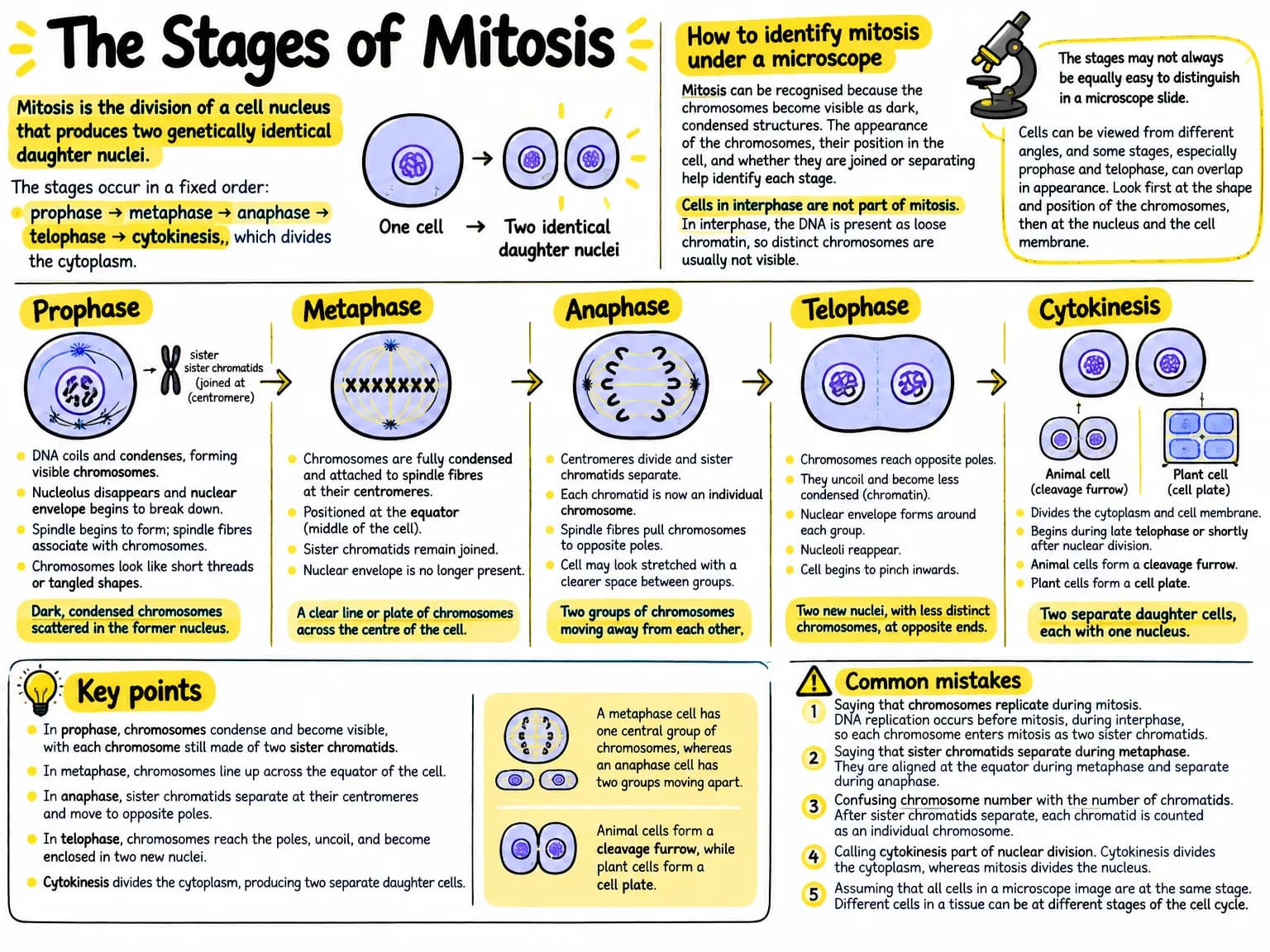
Mitosis is the division of one nucleus into two genetically identical nuclei. The main stages are prophase, metaphase, anaphase and telophase, followed by cytokinesis, which divides the cytoplasm and produces two daughter cells.
The Stages of Mitosis at a glance
Listen: The Stages of Mitosis
Two hosts talking it through, 2:58. The full transcript is below, so you can read along or skip the audio.
Transcript
AnnaWhen we talk about mitosis, what exactly is dividing?
MarcoOne nucleus divides into two genetically identical nuclei. Then cytokinesis divides the cytoplasm, producing two daughter cells. The usual order is prophase, metaphase, anaphase, telophase, and then cytokinesis.
AnnaSo where does prophase begin?
MarcoThe DNA copied before mitosis coils and condenses into visible chromosomes. Each chromosome has two identical sister chromatids joined at a centromere. The nucleolus disappears, the spindle begins forming, and centrosomes move toward opposite poles.
AnnaWhat would we notice under a microscope?
MarcoDark, shortened, threadlike or rodlike chromosomes become visible, but they are scattered rather than lined up across the middle. Early on, the nuclear envelope may still be partly visible. Some descriptions call its later breakdown prometaphase.
AnnaThen what changes in metaphase?
MarcoSpindle fibres attach to kinetochores at the centromeres. They position the duplicated chromosomes along the equator, or middle, of the cell. The sister chromatids are still joined. That is important: they do not separate until anaphase.
AnnaIs the microscope clue simply a line of chromosomes?
MarcoExactly. Metaphase shows a clear line across the centre, often like small, thick rods or duplicated chromosomes arranged in a plate. The nuclear envelope is no longer present.
AnnaWhat happens when the cell enters anaphase?
MarcoThe centromeres divide, and the sister chromatids separate. Each chromatid is now an individual daughter chromosome. Spindle fibres shorten and pull the daughter chromosomes toward opposite poles.
AnnaAnd how can we recognise that movement?
MarcoThere are two groups moving away from each other. The chromosomes may look like V shapes or other bent shapes, because their centromeres lead the way. They are not yet enclosed in new nuclei.
AnnaWhat makes telophase different?
MarcoThe chromosomes reach opposite poles and begin to uncoil into less distinct chromatin. A new nuclear envelope forms around each group, nucleoli reappear, and the spindle breaks down. Under a microscope, two groups sit at opposite ends, and two nuclei may be forming.
AnnaDoes the cell already split at that point?
MarcoNot completely. Cytokinesis divides the cytoplasm. In an animal cell, a contractile ring creates a cleavage furrow that pinches inward. In a plant cell, vesicles gather in the centre and form a cell plate, which develops into a new cell wall.
AnnaSo how do we avoid confusing cytokinesis with anaphase or telophase?
MarcoLook for the nuclei or chromosome groups. In cytokinesis, the nuclei have already reformed, and the cell is physically separating. In anaphase, chromosomes are still moving apart. The key sequence is condensing, lining up, separating, forming two nuclei, and then splitting the cytoplasm. Interphase comes before mitosis, but it is not one of its stages.
The notes
Prophase
During prophase, the DNA that was copied before mitosis coils and condenses into visible chromosomes. Each chromosome consists of two identical sister chromatids joined at a centromere. The nucleolus disappears, the spindle begins to form, and the centrosomes move towards opposite poles of the cell.
Under a microscope, prophase cells have dark, shortened, threadlike or rodlike chromosomes that are becoming visible, but the chromosomes are not arranged across the middle of the cell. The nuclear envelope may still be partly visible at early prophase. In some descriptions, the later breakdown of the nuclear envelope is called prometaphase rather than prophase.
Metaphase
During metaphase, spindle fibres attach to structures at the centromeres called kinetochores. The spindle fibres position the duplicated chromosomes along the equator, or middle, of the cell. The sister chromatids are still joined together at this stage.
Under a microscope, metaphase is recognised by the chromosomes forming a clear line across the centre of the cell. They often look like small, thick rods or duplicated chromosomes arranged in a plate. The nuclear envelope is no longer present.
Anaphase
During anaphase, the centromeres divide and the sister chromatids separate. Each chromatid is now considered an individual daughter chromosome. Spindle fibres shorten and pull the daughter chromosomes towards opposite poles of the cell.
Under a microscope, anaphase cells show two groups of chromosomes moving away from each other. The chromosomes may look like V shapes or other bent shapes because their centromeres lead as they are pulled towards the poles. The groups are not yet enclosed in new nuclei.
Telophase
During telophase, the chromosomes reach opposite poles and begin to uncoil, becoming less distinct as chromatin. A new nuclear envelope forms around each group of chromosomes, and nucleoli reappear. The spindle breaks down.
Under a microscope, telophase is recognised by two groups of chromosomes at opposite ends of the cell. These groups may look less dark and less sharply defined than the chromosomes in metaphase or anaphase. Two new nuclei can often be seen forming, and the cell may begin to pinch in the middle.
Cytokinesis
Cytokinesis is the division of the cytoplasm. In animal cells, a contractile ring causes a cleavage furrow to form, which deepens until the cell splits into two daughter cells. In plant cells, vesicles collect at the centre and form a cell plate, which develops into a new cell wall between the daughter cells.
Under a microscope, cytokinesis in an animal cell appears as a narrowing or pinching at the cell surface. In a plant cell, it appears as a developing partition across the middle. The nuclei have already reformed, so cytokinesis can be distinguished from anaphase by the presence of two nuclei or two chromosome groups in the new daughter cells.
Identifying the stages in order
The stages can be identified by following the position and appearance of the chromosomes. In prophase, chromosomes condense but are scattered. In metaphase, duplicated chromosomes line up at the equator. In anaphase, sister chromatids have separated into two moving groups. In telophase, two nuclei form and the chromosomes uncoil. Cytokinesis then completes the physical separation of the cells.
Microscope images are not always perfectly clear, and cells may not show every stage equally well. The key distinction is whether chromosomes are condensing, lined up, separating, or enclosed in two new nuclei. Interphase is not part of mitosis, although it is often shown in diagrams because the cell grows and copies its DNA before mitosis begins.
What to remember
- In prophase, copied DNA condenses into visible chromosomes, each made of two sister chromatids.
- In metaphase, duplicated chromosomes line up across the equator of the cell.
- In anaphase, centromeres divide and sister chromatids become separate daughter chromosomes.
- In telophase, chromosomes reach opposite poles, uncoil and become enclosed in two new nuclei.
- Cytokinesis divides the cytoplasm and produces two daughter cells.
- Animal cells form a cleavage furrow, while plant cells form a cell plate.
- The stage order is prophase, metaphase, anaphase, telophase, then cytokinesis.
The Stages of Mitosis as a mind map
- Stages of Mitosis
- Purpose and Sequence
- Nuclear division into two genetically identical nuclei
- Prophase → Metaphase → Anaphase → Telophase → Cytokinesis
- Interphase: DNA copying before mitosis, not part of mitosis
- Mitosis Stages
- Prophase
- Copied DNA condenses into visible chromosomes
- Each chromosome has two sister chromatids joined at a centromere
- Nucleolus disappears; spindle forms; centrosomes move to opposite poles
- Chromosomes are condensed and scattered, not aligned at the equator
- Metaphase
- Spindle fibres attach to kinetochores at centromeres
- Duplicated chromosomes line up at the cell equator
- Sister chromatids remain joined
- Anaphase
- Centromeres divide and sister chromatids separate
- Separated chromatids become daughter chromosomes
- Spindle fibres pull chromosomes toward opposite poles
- Telophase
- Chromosomes reach opposite poles and uncoil into chromatin
- New nuclear envelopes and nucleoli form
- Spindle breaks down
- Prophase
- Cytokinesis
- Division of the cytoplasm into two daughter cells
- Animal cells: contractile ring forms a cleavage furrow
- Plant cells: vesicles form a cell plate and new cell wall
- Stage Identification
- Prophase: condensing, scattered chromosomes
- Metaphase: chromosomes aligned across the centre
- Anaphase: two chromosome groups moving apart
- Telophase: two nuclei forming around uncoiled chromosomes
- Cytokinesis: physical separation of daughter cells
- Common Distinctions and Mistakes
- Chromatids separate in anaphase, not metaphase
- Chromosomes align at the equator in metaphase, not prophase
- Telophase reforms nuclei; cytokinesis divides the cytoplasm
- Plant cells form a cell plate, not a cleavage furrow
- Purpose and Sequence
Flashcards
- What is mitosis?
- Mitosis is the division of one nucleus into two genetically identical nuclei.
- What is the correct order of the stages of mitosis?
- Prophase, metaphase, anaphase, telophase, followed by cytokinesis.
- What happens during prophase?
- Copied DNA condenses into visible chromosomes, each consisting of two sister chromatids joined at a centromere. The spindle begins to form and centrosomes move toward opposite poles.
- How can prophase be identified under a microscope?
- Chromosomes appear dark, shortened, and threadlike or rodlike, but they are scattered rather than lined up across the cell's middle.
- What happens during metaphase?
- Spindle fibres attach to kinetochores, and duplicated chromosomes line up along the equator of the cell. Sister chromatids are still joined.
- How can metaphase be identified under a microscope?
- Chromosomes form a clear line or plate across the centre of the cell, and the nuclear envelope is absent.
- What happens during anaphase?
- Centromeres divide, causing sister chromatids to separate and become individual daughter chromosomes. Spindle fibres pull them toward opposite poles.
- How can anaphase be identified under a microscope?
- Two groups of chromosomes are moving away from each other, often appearing V-shaped or bent, and they are not yet enclosed in new nuclei.
- What happens during telophase?
- Chromosomes reach opposite poles, uncoil into chromatin, and become enclosed by new nuclear envelopes. Nucleoli reappear and the spindle breaks down.
- How can telophase be identified under a microscope?
- Two less-distinct chromosome groups or two newly forming nuclei appear at opposite ends of the cell; the cell may begin to pinch in the middle.
- What is cytokinesis, and how does it differ in animal and plant cells?
- Cytokinesis divides the cytoplasm to produce two daughter cells. Animal cells form a cleavage furrow, while plant cells form a cell plate that develops into a new cell wall.
- How can cytokinesis be distinguished from anaphase?
- During cytokinesis, nuclei have already reformed and the cell is physically separating. During anaphase, chromosomes are still moving toward opposite poles and are not enclosed in nuclei.
Test yourself
During which stage do sister chromatids become individual daughter chromosomes?
Sister chromatids separate when the centromeres divide during anaphase.
Which microscopic feature most clearly identifies metaphase?
In metaphase, duplicated chromosomes line up across the equator of the cell.
Which event is characteristic of telophase rather than cytokinesis?
Telophase involves the formation of new nuclear envelopes around the chromosome groups.
How does cytokinesis usually occur in a plant cell?
Plant cytokinesis uses vesicles that gather centrally to form a cell plate and new cell wall.
A cell shows two chromosome groups moving toward opposite poles, with no new nuclei yet visible. Which stage is it in?
Anaphase is identified by separated chromosomes moving toward opposite poles before new nuclei form.
Common mistakes
- Saying that sister chromatids separate during metaphase, when they remain joined until anaphase.
- Calling the two chromatids of one duplicated chromosome two separate chromosomes before the centromere divides.
- Confusing cytokinesis with telophase, since telophase reforms the nuclei while cytokinesis divides the cytoplasm.
- Thinking that chromosomes line up at the cell equator during prophase, when this happens during metaphase.
- Forgetting that plant cells form a cell plate rather than using a cleavage furrow.
Make this for your own notes
Everything on this page came from one source, generated in about a minute. Paste a chapter, upload a PDF or photograph your notes and get the same set back.
Try it freeNo credit card. Free every month.