
Mitosis vs Meiosis — Visual Diagram & Comparison
Cell division made clear — the phases of mitosis and the key mitosis-vs-meiosis differences — in one comparison diagram.
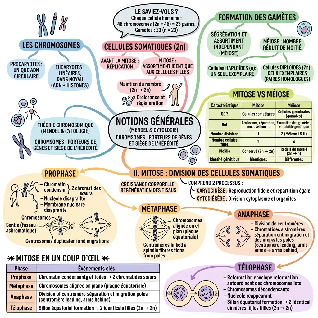
Early in the 1900s, Mendel's experiments combined with cytological evidence from mitosis and meiosis led to the chromosome theory of heredity: chromosomes are the cellular structures that carry genes. In prokaryotes the chromosome is a single circular DNA molecule; in eukaryotes chromosomes are linear, held in the nucleus and bound to histone proteins. Somatic cells are diploid (2n) and replicate their chromosomes before mitosis so each daughter cell is identical to the parent. Mitosis divides somatic cells, keeps the chromosome number constant (2n to 2n) and drives growth and regeneration, through four phases: prophase, metaphase, anaphase and telophase. Meiosis divides germ cells, halves the chromosome number (2n to n) through two divisions, forms gametes and generates genetic variability. Human cells carry 46 chromosomes; gametes carry 23.
What's in this visual
Mitosis and meiosis are a classic exam pairing — easy to confuse, and usually revised from a wall of phase descriptions and a comparison table. The diagram above takes the same content and lays it out side by side, so the four phases of mitosis read as a sequence and the mitosis-versus-meiosis contrast is visible at a glance. Here is the full breakdown.
The chromosome theory of heredity
In the early 1900s, Mendel's inheritance experiments combined with cytological observations of cell division to produce the chromosome theory of heredity: chromosomes are the cellular structures that carry genes and are the physical seat of inheritance. Chromosomes differ between cell types — in prokaryotes they are a single circular DNA molecule loosely associated with protein, while in eukaryotes they are linear, contained in the nucleus and tightly bound to histone proteins. This framing matters because mitosis and meiosis are simply the two ways those chromosomes are passed on.
Mitosis: division of somatic cells
Mitosis is the division of somatic cells — the body's non-reproductive cells. It drives bodily growth and the regeneration of tissues, and it keeps the chromosome number constant: a diploid (2n) parent cell yields two genetically identical diploid daughter cells. Mitosis involves two coordinated processes: karyokinesis, the faithful copying and equal distribution of chromosomes, and cytokinesis, the division of the cytoplasm and its organelles. Before mitosis begins, the cell replicates its chromosomes so that each is made of two sister chromatids ready to be separated.
The four phases of mitosis
Mitosis runs through four phases. In prophase, chromatin condenses into visible chromosomes of two sister chromatids, the nucleolus and nuclear membrane disappear, and the spindle forms as centrosomes migrate to opposite poles. In metaphase, chromosomes align on the equatorial plate, their centromeres attached to spindle fibres from both poles. In anaphase, centromeres split and sister chromatids are pulled to opposite poles, centromere first. In telophase, nuclear envelopes reform, chromosomes unwind back into chromatin, the nucleolus reappears, and cytokinesis cleaves the cell into two identical daughter cells.
Meiosis and why it halves the chromosome number
Meiosis is the division of germ cells in the gonads, and its purpose is to produce gametes. Unlike mitosis, it involves two successive divisions — meiosis I and meiosis II — which together turn one diploid cell into four cells, each haploid (n) with half the original chromosome number. The independent segregation and assortment of chromosomes during meiosis is also the source of genetic variability, so the daughter cells are not identical to one another. In humans, this is what reduces the 46 chromosomes of a body cell to the 23 carried by each sperm or egg.
Mitosis vs meiosis — the key differences
The two processes are best learned by direct contrast. Mitosis occurs in somatic cells; meiosis in germ cells. Mitosis serves growth, repair and renewal; meiosis serves gamete formation and genetic variability. Mitosis is a single division producing two daughter cells; meiosis is two divisions producing four. Mitosis conserves ploidy (2n to 2n); meiosis halves it (2n to n). And mitosis yields genetically identical daughter cells, while meiosis yields genetically different ones. These paired rows are exactly what a side-by-side visual is built to hold.
Why comparison topics need a side-by-side visual
Comparison topics fail in plain notes because the two halves end up on different pages, and you re-read both every time to reconstruct the contrast. A side-by-side visual fixes mitosis and meiosis in adjacent columns, so each difference — cell type, number of divisions, daughter-cell count, ploidy, genetic identity — lines up on a single row. You stop memorising two separate descriptions and start seeing one comparison, which is exactly how exam questions are framed.
For teachers
The problem
- Students consistently confuse mitosis and meiosis because notes describe them in separate sections rather than side by side.
- The four phases of mitosis are a sequence, but a flat list of phase descriptions hides the order and the events.
- Drawing a clean, parallel comparison on the board takes time and rarely fits legibly.
How to use it in class
- Hand it out as a revision sheet before the cell division test.
- Project the four phases and walk through prophase to telophase as a sequence.
- Use the side-by-side columns to drill the mitosis-versus-meiosis contrast row by row.
- Blank the comparison table cells to turn the diagram into a quick recall quiz.
For students & visual learners
The problem
- You can describe the phases of mitosis but mix up which features belong to meiosis.
- Prophase, metaphase, anaphase and telophase blur together when revised as a paragraph.
- The mitosis-versus-meiosis differences feel like a list to memorise rather than a pattern to understand.
How to use it to study
- Revise both processes in one glance instead of flipping between two sets of notes.
- Use the phase sequence to recall prophase to telophase in the correct order.
- Read the comparison columns row by row so each difference sticks as a pair.
- Pin it above your desk so the contrast becomes automatic before the exam.
Make your own visual like this
Paste your notes or upload a PDF
Drop in your own class notes, a textbook chapter or a PDF. Any subject, any language.
Pick a visual style
Choose a sketchnote, infographic or diagram style that fits the topic.
Generate and download
In about 15–40 seconds you get a one-page visual you can print, share or revise from.
Frequently asked questions
What are the four phases of mitosis?
The four phases of mitosis are prophase (chromosomes condense, the spindle forms), metaphase (chromosomes align on the equatorial plate), anaphase (sister chromatids separate to opposite poles) and telophase (nuclear envelopes reform and the cell divides into two identical daughter cells).
What is the main difference between mitosis and meiosis?
Mitosis is a single division of somatic cells that produces two genetically identical diploid daughter cells and conserves the chromosome number. Meiosis is two divisions of germ cells that produce four genetically different haploid gametes and halves the chromosome number (2n to n).
Does VisualNote AI work with notes in French or other languages?
Yes. The source notes for this very diagram were written in French, and the page was generated in English — VisualNote AI handles many languages and can translate as it summarises. Paste notes in any language and get a clear visual.
More visual examples


Turn your notes into a visual
Paste any notes or upload a PDF and get a sketchnote-style visual in under a minute.