
Heart Anatomy & the Cardiac Cycle: Diagram, Notes and Flashcards
Six pages of cardiology lecture notes, rebuilt as a single labelled heart diagram you can read in one pass. The same source also produced a flashcard deck, so you can look at the structure and then test yourself on it.
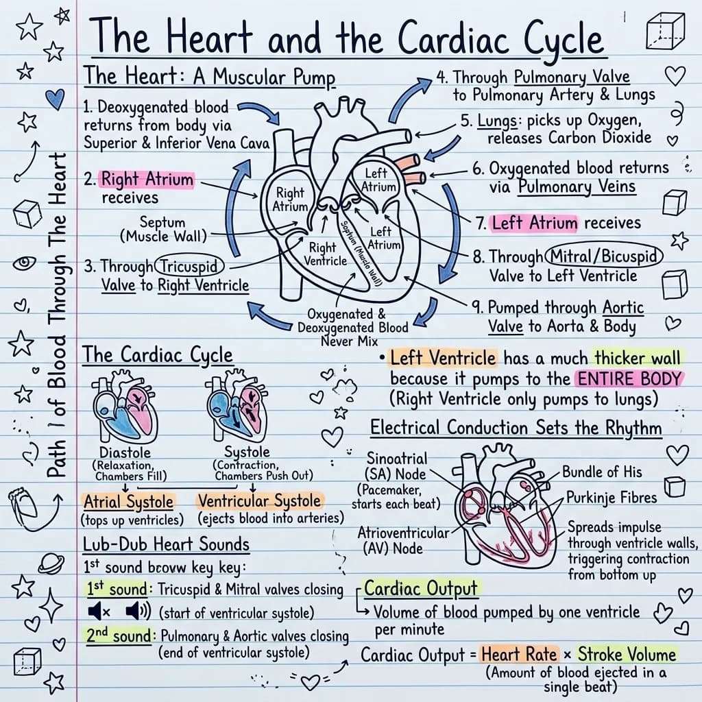
2.1 Gross structure of the heart. The heart is a four chambered muscular pump lying in the mediastinum, divided by the interatrial and interventricular septa into a right side that handles deoxygenated blood and a left side that handles oxygenated blood. The two atria are thin walled receiving chambers and the two ventricles are thick walled pumping chambers; the left ventricular wall is roughly three times thicker than the right because it ejects blood into the systemic circulation against a much higher afterload, while the right ventricle ejects into the low pressure pulmonary circulation. 2.2 Valves. Four valves enforce one way flow: the tricuspid valve between right atrium and right ventricle, the pulmonary valve at the origin of the pulmonary trunk, the mitral or bicuspid valve between left atrium and left ventricle, and the aortic valve at the origin of the aorta. 2.3 Circulation. Deoxygenated blood returns via the superior and inferior venae cavae to the right atrium, passes the tricuspid valve into the right ventricle, is ejected through the pulmonary valve into the pulmonary arteries to the lungs, returns oxygenated through the pulmonary veins to the left atrium, passes the mitral valve into the left ventricle and is ejected through the aortic valve into the aorta. 2.4 The cardiac cycle. Diastole is the relaxation and filling phase and systole is the contraction and ejection phase; the first heart sound S1, described as lub, results from closure of the atrioventricular valves at the onset of ventricular systole and the second heart sound S2, described as dub, results from closure of the semilunar valves at the onset of diastole. 2.5 Conduction. The sinoatrial node in the wall of the right atrium acts as the pacemaker, the impulse spreads across the atria to the atrioventricular node where it is briefly delayed, then travels down the bundle of His, into the left and right bundle branches and out through the Purkinje fibres to depolarise the ventricular myocardium from the apex upward. 2.6 Output. Cardiac output is the product of heart rate and stroke volume and is normally about five litres per minute at rest.
The flashcards from the same notes
Exports as Anki CSV, printable PDF, or plain text.
Which valve sits between the right atrium and the right ventricle?
The tricuspid valve, named for its three cusps.
What produces the first heart sound, S1 or lub?
Closure of the atrioventricular valves, the tricuspid and the mitral, at the start of ventricular systole.
Why is the wall of the left ventricle thicker than the wall of the right?
The left ventricle pumps blood through the whole systemic circulation against high resistance, while the right ventricle only pushes blood to the nearby lungs at low pressure.
Which structure is the natural pacemaker of the heart, and where is it?
The sinoatrial (SA) node, in the wall of the right atrium.
Why does the AV node delay the impulse before passing it on?
The delay gives the atria time to finish emptying into the ventricles before ventricular contraction begins.
State the formula for cardiac output.
Cardiac output = heart rate x stroke volume, about 5 litres per minute at rest.
Make a deck from your own notes with the AI flashcard generator.
What's in this visual
The heart is the topic where almost everyone can name the parts and almost nobody can trace the flow. Chambers, valves, sounds, conduction and output are usually taught as five separate lists, when they are really five views of the same pump. The diagram above puts them on one page so the relationships are visible; the flashcards underneath then check whether the relationships actually stuck. Here is the full breakdown.
Four chambers, and why the left ventricle is the thick one
The heart is two pumps sharing a wall. The right atrium receives deoxygenated blood from the body and the right ventricle sends it to the lungs. The left atrium receives oxygenated blood from the lungs and the left ventricle sends it to the whole body. Atria are thin walled because they only need to nudge blood downward into the ventricles. The left ventricular wall is roughly three times thicker than the right for one reason: it pushes against systemic resistance, while the right ventricle only has to reach the lungs sitting a few centimetres away at low pressure. Once you attach that reason to the picture, the thickness stops being a fact to memorise.
The four valves and the full path of blood
Valves exist to stop backflow, and the order they appear in is the order blood meets them. Body to superior and inferior venae cavae, into the right atrium, through the tricuspid valve into the right ventricle, out through the pulmonary valve into the pulmonary arteries and to the lungs. Back through the pulmonary veins into the left atrium, through the mitral (bicuspid) valve into the left ventricle, and out through the aortic valve into the aorta. Two memory hooks are worth keeping: the tricuspid has three cusps and sits on the right; the pulmonary arteries are the only arteries carrying deoxygenated blood, and the pulmonary veins the only veins carrying oxygenated blood.
Diastole, systole and the lub-dub
The cardiac cycle has two phases. Diastole is relaxation and filling; the ventricles are passive and blood pours in from the atria. Systole is contraction and ejection. The two heart sounds simply mark the boundaries. S1 (lub) is the atrioventricular valves, tricuspid and mitral, slamming shut as the ventricles begin to contract. S2 (dub) is the semilunar valves, pulmonary and aortic, snapping shut as the ventricles relax. The sounds are valves closing, never valves opening: that single sentence answers a surprising number of exam questions.
The conduction system that sets the rhythm
Electrical activity runs ahead of mechanical activity. The SA node in the right atrial wall fires on its own and sets the pace. The impulse spreads across both atria, which contract, and reaches the AV node, where it is deliberately delayed for a fraction of a second so the atria can finish emptying before the ventricles fire. It then runs down the bundle of His, splits into the left and right bundle branches, and fans out through the Purkinje fibres, which depolarise the ventricles from the apex upward so blood is squeezed toward the outflow valves rather than away from them. Every rhythm strip you will ever read is this pathway working or failing at a specific point.
Cardiac output, and why a visual plus a deck beats re-reading
Cardiac output is the summary number: CO = heart rate x stroke volume, roughly 70 beats per minute times 70 millilitres, giving about five litres per minute at rest. Anything that changes rate or filling changes output, which is why the formula sits at the centre of so much cardiology. The diagram above holds the whole structure in one field of view; the six flashcards below were generated from the exact same lecture notes, so the questions test the same relationships the picture shows instead of drifting off into unrelated trivia. That pairing is the point of flashcards for students and teachers: look at the structure, then prove you can produce it from memory.
For teachers
The problem
- Blood flow, valves and conduction are three separate diagrams in most textbooks, so students never see them as one system.
- Drawing an accurate heart on a whiteboard eats fifteen minutes of a fifty minute class.
- Students arrive able to name chambers but unable to explain why the left ventricle is thicker.
How to use it in class
- Project the diagram and trace the path of blood with a pointer while the class follows.
- Print it as a revision sheet before the cardiovascular assessment.
- Blank the valve labels and use it as a five minute recall starter.
- Set the flashcard deck as homework and open the next lesson with the two cards most students missed.
For students & visual learners
The problem
- You can list the four valves but stall halfway through tracing blood from the vena cava to the aorta.
- S1 and S2 blur together, and you can never remember whether the sound is opening or closing.
- The conduction pathway is five names in a row with nothing to hang them on.
How to use it to study
- Follow the arrows once a day until the path of blood becomes automatic.
- Use the diagram to answer, then the flashcards to check you were not just recognising the layout.
- Keep it open next to practice ECG questions so the SA node to Purkinje route stays in view.
- Revise the whole cardiac cycle in two minutes before a placement shift or an OSCE station.
Make your own visual like this
Paste your notes or upload a PDF
Drop in your own class notes, a textbook chapter or a PDF. Any subject, any language.
Pick a visual style
Choose a sketchnote, infographic or diagram style that fits the topic.
Generate and download
In about 15–40 seconds you get a one-page visual you can print, share or revise from.
Frequently asked questions
What is the correct order of blood flow through the heart?
Venae cavae, right atrium, tricuspid valve, right ventricle, pulmonary valve, pulmonary arteries, lungs, pulmonary veins, left atrium, mitral valve, left ventricle, aortic valve, aorta.
What is the difference between diastole and systole?
Diastole is the relaxation phase, when the ventricles fill with blood. Systole is the contraction phase, when the ventricles eject blood into the pulmonary artery and the aorta.
Can I turn my own anatomy notes into a diagram and a deck like this?
Yes. Paste your lecture notes or upload a PDF and you get a labelled visual plus a matching set of cards from the same source. Try the AI flashcard generator.
More visual examples

Turn your notes into a visual
Paste any notes or upload a PDF and get a sketchnote-style visual in under a minute.