
The Respiratory System: Labelled Diagram, Notes and Flashcards
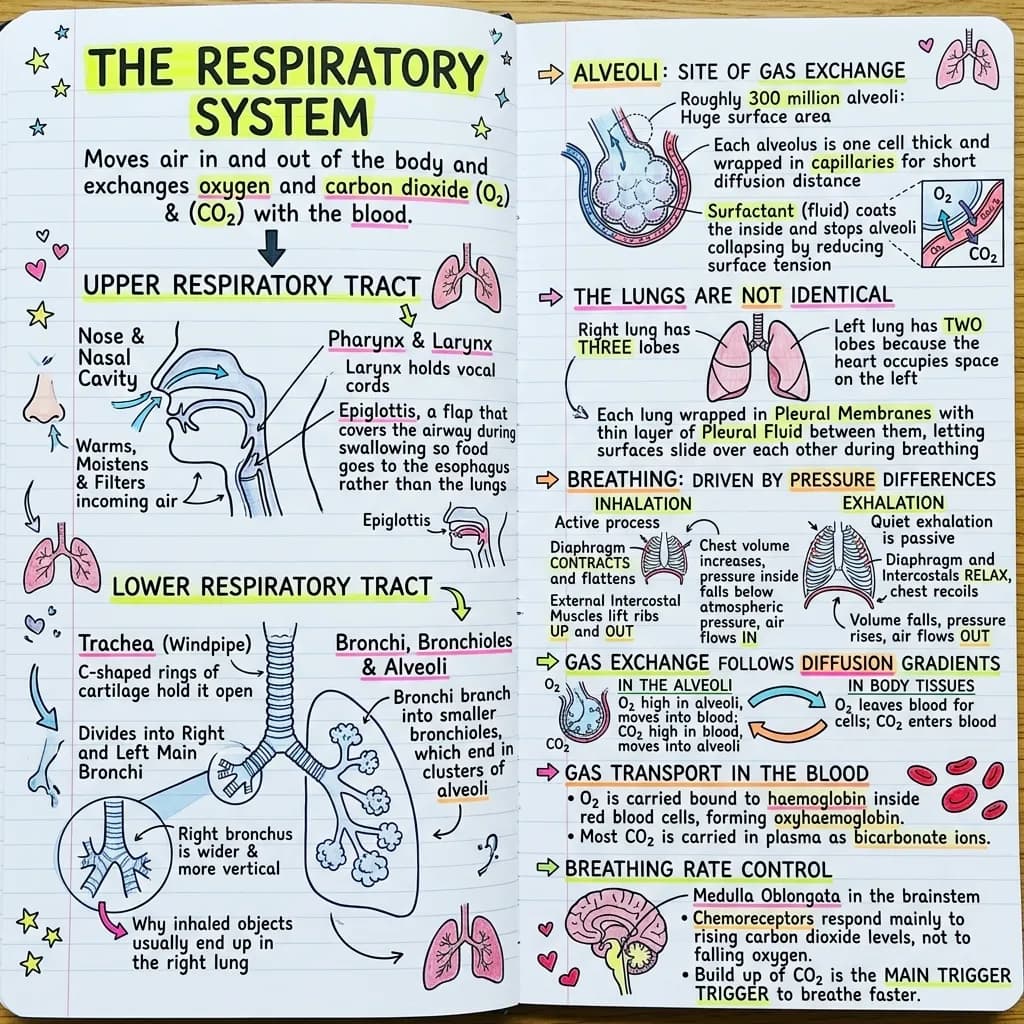
A chapter of respiratory notes turned into a single labelled diagram that runs from the nose to the alveolus without a page turn. The flashcard deck was built from the same chapter, so the questions match the picture exactly.
Chapter 9: the respiratory system. The respiratory tract is divided into an upper tract comprising the nose, nasal cavity, pharynx and larynx, and a lower tract comprising the trachea, bronchi, bronchioles and alveoli within the lungs. During swallowing the epiglottis, a flap of elastic cartilage, folds over the laryngeal opening so that food passes into the oesophagus rather than the airway. The trachea is held open by approximately sixteen to twenty C shaped rings of hyaline cartilage that are incomplete posteriorly, the gap being bridged by smooth muscle so that the oesophagus can expand during swallowing. The trachea bifurcates at the carina into the right and left main bronchi; the right main bronchus is wider, shorter and more vertical than the left, and inhaled foreign objects therefore lodge in it preferentially. The right lung has three lobes and the left lung has two, the reduced number on the left reflecting the space occupied by the heart at the cardiac notch. Gas exchange occurs in approximately three hundred million alveoli, each lined by a single layer of squamous epithelium and coated internally by surfactant, a phospholipid that lowers surface tension and prevents alveolar collapse at the end of expiration; deficiency in premature infants produces respiratory distress syndrome. Inhalation is an active process in which the diaphragm contracts and flattens while the external intercostal muscles elevate the ribs, increasing thoracic volume, reducing intrapulmonary pressure below atmospheric pressure and drawing air inward; quiet exhalation is passive and results from elastic recoil of the lungs and chest wall. Gas exchange occurs by simple diffusion down partial pressure gradients across the respiratory membrane. Oxygen is transported almost entirely bound to haemoglobin as oxyhaemoglobin, whereas carbon dioxide is transported mainly as bicarbonate ion in plasma, with smaller fractions as carbaminohaemoglobin and dissolved gas. Respiratory rate is regulated principally by central chemoreceptors responding to rising arterial carbon dioxide and hydrogen ion concentration rather than to falling oxygen.
The flashcards from the same notes
Exports as Anki CSV, printable PDF, or plain text.
Why do inhaled objects usually lodge in the right main bronchus?
The right main bronchus is wider, shorter and more vertical than the left, so objects fall into it more easily.
Why are the cartilage rings of the trachea C-shaped rather than complete?
The gap at the back lets the oesophagus expand during swallowing, while the cartilage in front keeps the airway permanently open.
What does surfactant do in the alveoli?
It lowers surface tension so the alveoli do not collapse at the end of exhalation.
Why does the right lung have three lobes and the left only two?
The heart sits slightly to the left, occupying space at the cardiac notch, so the left lung has room for only two lobes.
Which muscle action makes inhalation an active process?
The diaphragm contracts and flattens while the external intercostals raise the ribs, increasing thoracic volume and dropping the pressure inside the lungs below atmospheric.
Which gas is the main driver of breathing rate?
Carbon dioxide. Central chemoreceptors respond to rising carbon dioxide and hydrogen ion levels, not to falling oxygen.
Make a deck from your own notes with the AI flashcard generator.
What's in this visual
Breathing is easy to describe and hard to explain. Most students can list the structures from nose to lung but stall on the questions that matter: why objects go right, why exhaling costs nothing, why carbon dioxide and not oxygen sets your breathing rate. The diagram above runs the whole tract in one image with the reasons attached to the parts. Here is the walkthrough.
Upper tract, lower tract, and the flap that keeps them separate
The upper respiratory tract is the nose, nasal cavity, pharynx and larynx: its job is to warm, humidify and filter incoming air. The lower respiratory tract is the trachea, bronchi, bronchioles and alveoli, where conduction ends and exchange begins. The pharynx is shared with the digestive tract, which is a design problem solved by the epiglottis: a flap of elastic cartilage that folds over the laryngeal opening every time you swallow, so food is directed into the oesophagus. When that reflex fails, material enters the airway and you get aspiration, which is why the epiglottis appears in clinical questions far more often than its size suggests.
Why the trachea has C-shaped rings and why objects go right
The trachea is held permanently open by around sixteen to twenty rings of hyaline cartilage, and those rings are C-shaped rather than complete for a specific reason: the gap sits at the back, bridged by soft muscle, so the oesophagus can bulge forward as a swallowed bolus passes. Rigid airway at the front, flexibility at the back. Below the carina the trachea splits, and the right main bronchus is wider, shorter and more vertical than the left. Gravity and geometry do the rest, so an inhaled peanut, tooth or tablet lands in the right bronchus far more often than the left. That single asymmetry explains where aspiration pneumonia tends to appear on a chest film.
Alveoli, surfactant, and the lung that gave up a lobe
Around three hundred million alveoli give the lungs an exchange surface roughly the size of a tennis court, each one a single squamous cell thick and wrapped in capillaries. Their internal coating of surfactant lowers surface tension so the alveoli do not collapse shut at the end of every breath; premature babies who have not yet produced enough of it develop respiratory distress syndrome, which is the clinical proof of what surfactant is for. Higher up, the asymmetry of the lungs has a simple cause: the right lung has three lobes and the left has two, because the heart sits slightly left and takes up the space at the cardiac notch.
Breathing in costs energy, breathing out usually does not
Inhalation is active. The diaphragm contracts and flattens, the external intercostals lift the ribs up and out, thoracic volume increases, pressure inside the lungs drops below atmospheric, and air moves in down the pressure gradient. Quiet exhalation is passive. The muscles simply relax and the elastic recoil of lung tissue and chest wall pushes air back out; no contraction is needed until you are exercising or forcing air out. Once you frame breathing as a pressure story rather than a muscle list, the diagram becomes self explanatory: volume up means pressure down means air in.
Gas exchange, transport, and the gas that actually drives your breathing
Exchange itself is passive: oxygen and carbon dioxide diffuse across the respiratory membrane down their partial pressure gradients, no pumping involved. Transport differs by gas. Oxygen travels almost entirely bound to haemoglobin as oxyhaemoglobin, while most carbon dioxide is carried in plasma as bicarbonate, with smaller amounts on haemoglobin and dissolved in solution. The counterintuitive part is control: your breathing rate is set mainly by rising carbon dioxide detected by central chemoreceptors, not by falling oxygen, which only takes over as a stimulus at severely low levels. That is the fact most decks skip, so it was deliberately kept in the six cards below, generated from the same chapter as the diagram. If you want the same pairing from your own notes, start with the AI flashcard generator.
For teachers
The problem
- The tract, the mechanics and the gas transport chemistry usually live in three different diagrams.
- Students memorise the structures but cannot explain why exhalation is passive.
- The carbon dioxide drive for breathing is counterintuitive and gets taught in a single sentence that nobody retains.
How to use it in class
- Use the pathway image as a lesson opener and add one mechanism per slide.
- Print it as a labelled and unlabelled pair for a structure naming exercise.
- Anchor a practical on breathing rate to the chemoreceptor panel.
- Set the deck as pre-reading so the class arrives already holding the vocabulary.
For students & visual learners
The problem
- You can name the parts but freeze when asked why an inhaled object goes right.
- Inhalation and exhalation get described in muscle names rather than pressure changes, so nothing sticks.
- Oxygen on haemoglobin and carbon dioxide as bicarbonate get swapped under exam pressure.
How to use it to study
- Trace the airway once a day until nose to alveolus is automatic.
- Use the pressure arrows to answer breathing mechanics questions without memorising sentences.
- Test yourself with the six cards, then check any miss against the diagram.
- Keep it beside past papers so every wrong answer has a picture to go back to.
Make your own visual like this
Paste your notes or upload a PDF
Drop in your own class notes, a textbook chapter or a PDF. Any subject, any language.
Pick a visual style
Choose a sketchnote, infographic or diagram style that fits the topic.
Generate and download
In about 15–40 seconds you get a one-page visual you can print, share or revise from.
Frequently asked questions
What is the difference between the upper and lower respiratory tract?
The upper tract is the nose, nasal cavity, pharynx and larynx, and it conditions incoming air. The lower tract is the trachea, bronchi, bronchioles and alveoli, where conduction ends and gas exchange takes place.
Why is exhalation passive at rest?
Because the inspiratory muscles simply relax and the elastic recoil of the lungs and chest wall pushes air out. Exhalation only becomes active during exercise or forced breathing, when the abdominal and internal intercostal muscles are recruited.
Can I get a diagram and a flashcard deck from my own biology chapter?
Yes. Upload the chapter or paste your notes and both are produced from the same source, so the cards test what the diagram shows. Try the notes-to-visual generator.
More visual examples
Turn your notes into a visual
Paste any notes or upload a PDF and get a sketchnote-style visual in under a minute.