
The Nervous System: Neuron Diagram, Reflex Arc and Flashcards
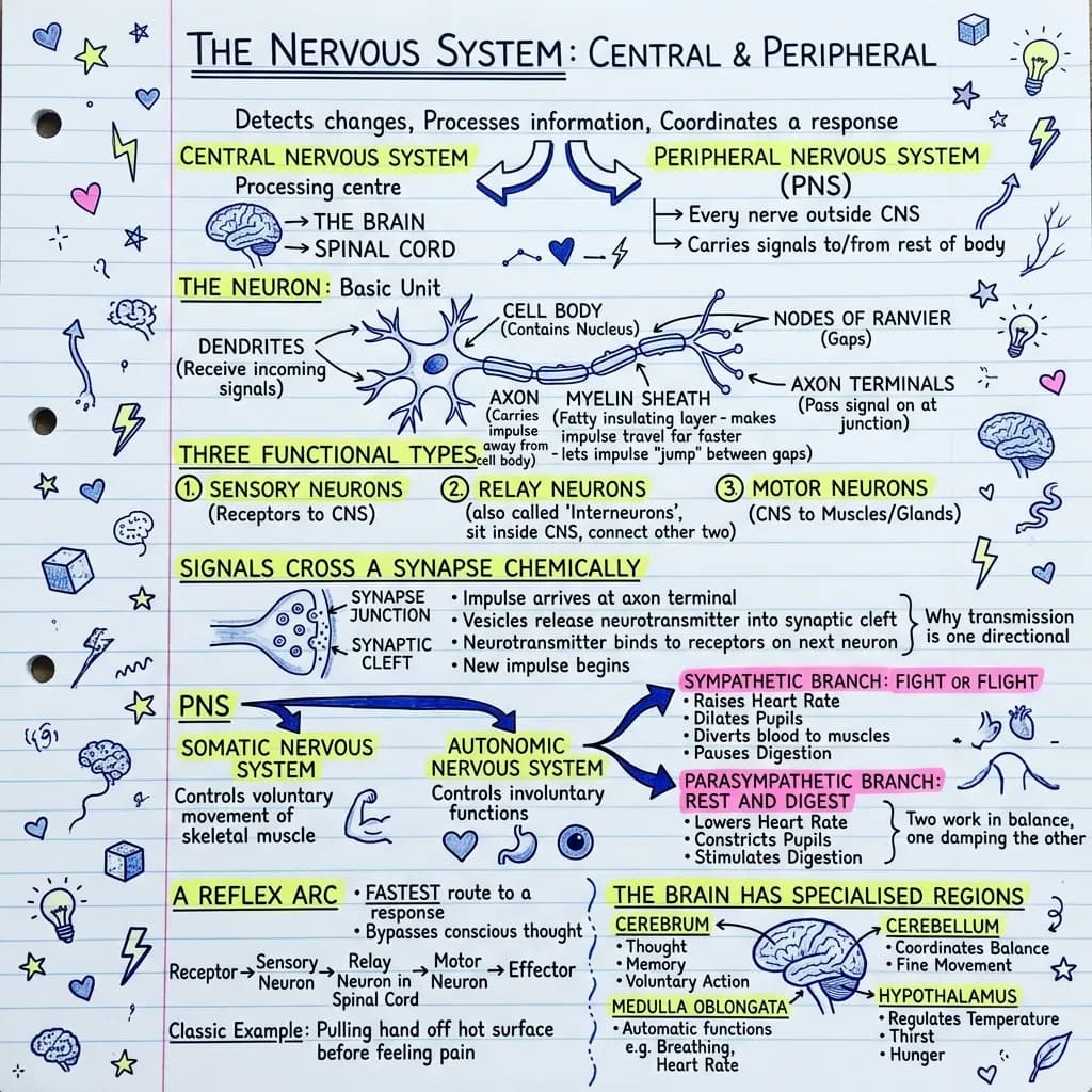
A full nervous system chapter compressed into one map that goes from a single neuron out to the whole brain. The six flashcards were generated from the same chapter, so revising the visual and testing yourself cover identical ground.
Unit 12: coordination and control. The nervous system is divided into the central nervous system, consisting of the brain and spinal cord, and the peripheral nervous system, consisting of all nerves outside those two structures. The functional unit is the neuron, which consists of dendrites that receive incoming signals, a cell body containing the nucleus, an axon that conducts the impulse away from the cell body, a myelin sheath formed by Schwann cells that insulates the axon, gaps in that sheath called nodes of Ranvier at which the impulse jumps in saltatory conduction, and axon terminals that form junctions with the next cell. Three functional types are recognised: sensory neurons carrying impulses from receptors to the central nervous system, relay neurons or interneurons lying within the central nervous system, and motor neurons carrying impulses from the central nervous system to effectors such as muscles and glands. Neurons do not touch; they are separated by a synaptic cleft across which transmission is chemical. An arriving impulse triggers vesicles in the presynaptic terminal to release neurotransmitter, which diffuses across the cleft and binds receptors on the postsynaptic membrane. Because vesicles are present only on the presynaptic side and receptors only on the postsynaptic side, transmission is unidirectional. The peripheral nervous system is subdivided into the somatic nervous system, which controls voluntary skeletal muscle, and the autonomic nervous system, which controls involuntary activity. The autonomic system is further divided into the sympathetic division, responsible for the fight or flight response, which increases heart rate, dilates pupils and airways and diverts blood to skeletal muscle, and the parasympathetic division, responsible for rest and digest activity, which slows heart rate, constricts pupils and stimulates digestion. A reflex arc allows a rapid involuntary response and follows the pathway stimulus, receptor, sensory neuron, relay neuron in the spinal cord, motor neuron, effector, response, bypassing conscious processing by the brain. Major brain regions include the cerebrum, responsible for thought, memory, reasoning and voluntary movement, the cerebellum, responsible for balance, posture and coordination of fine movement, the medulla oblongata, which regulates heart rate, breathing and blood pressure, and the hypothalamus, which regulates body temperature, thirst, hunger and hormone release through the pituitary gland.
The flashcards from the same notes
Exports as Anki CSV, printable PDF, or plain text.
Which two structures make up the central nervous system?
The brain and the spinal cord.
What do the myelin sheath and the nodes of Ranvier do?
The myelin sheath insulates the axon, and the impulse jumps from node to node in saltatory conduction, which greatly speeds transmission.
Why can a chemical synapse only transmit in one direction?
Neurotransmitter vesicles are found only in the presynaptic terminal and receptors only on the postsynaptic membrane, so the signal cannot cross the other way.
Name the three types of neuron in a reflex arc, in order.
Sensory neuron, then relay neuron (interneuron) in the spinal cord, then motor neuron.
Which autonomic division produces the fight or flight response?
The sympathetic division: it raises heart rate, dilates pupils and airways and diverts blood to skeletal muscle.
Which brain region controls heart rate and breathing?
The medulla oblongata.
Make a deck from your own notes with the AI flashcard generator.
What's in this visual
The nervous system is taught as one topic but it is really four: a cell, a junction, a circuit and a set of control centres. Students who struggle usually know all four separately and cannot connect them, which is exactly what a single map fixes. The visual above holds the neuron, the synapse, the reflex arc and the brain regions in one field of view. Here is each layer in turn.
CNS and PNS: the two halves of the system
The central nervous system is just two structures, the brain and the spinal cord, and it is where processing and decision making happen. The peripheral nervous system is everything else: every nerve running out to skin, muscle, gland and organ. The peripheral side splits again into the somatic division, which drives voluntary skeletal muscle, and the autonomic division, which runs everything you never think about. Getting this two by two structure clear first makes the rest of the topic fall into place, because every later term slots into one of these boxes.
Inside one neuron, and why myelin makes it fast
A neuron is a signal running in one direction across a specialised cell. Dendrites receive input and pass it to the cell body, which holds the nucleus. The axon carries the impulse away, wrapped in a fatty myelin sheath that insulates it. The sheath is not continuous: the gaps are the nodes of Ranvier, and the impulse jumps from node to node in saltatory conduction, which is far faster than crawling along the whole membrane. At the end, the axon terminals hand the signal over to the next cell. Damage to myelin slows or blocks conduction, which is why demyelinating disease produces such wide ranging symptoms from one underlying defect.
The synapse, and why signals only travel one way
Neurons never touch. They are separated by a tiny gap, the synaptic cleft, and the signal crosses it chemically. The arriving impulse makes vesicles in the presynaptic terminal fuse and release neurotransmitter, which diffuses across the gap and binds receptors on the postsynaptic membrane, starting a new impulse there. The reason transmission is one directional is structural and worth stating precisely: only the presynaptic side stores vesicles, and only the postsynaptic side carries receptors. Nothing prevents the chemistry from working backwards; the machinery simply is not there.
Three neuron types and the reflex arc they build
The three functional types are sensory neurons, which carry impulses from receptors inward, relay neurons (interneurons), which sit within the central nervous system, and motor neurons, which carry impulses outward to effectors. Put them in a line and you have the reflex arc: stimulus, receptor, sensory neuron, relay neuron in the spinal cord, motor neuron, effector, response. The whole point is that the loop closes in the spinal cord rather than the brain, which is why your hand is already off the hot pan before you consciously register the heat. Awareness arrives afterwards, as a separate signal.
Autonomic balance and the brain regions in charge
The autonomic system runs as an opposing pair. Sympathetic is fight or flight: heart rate up, pupils and airways dilated, blood diverted to skeletal muscle, digestion suspended. Parasympathetic is rest and digest, doing the reverse on each count, which makes the two easiest to learn as a mirrored list rather than two separate ones. Above them sit the control centres: the cerebrum for thought, memory and voluntary movement, the cerebellum for balance and fine coordination, the medulla oblongata for heart rate and breathing, and the hypothalamus for temperature, thirst, hunger and hormone release. The six cards below came from the same chapter as this map, so they check the connections rather than isolated definitions; the same pairing is available for any chapter through flashcards for students and teachers.
For teachers
The problem
- Neuron structure, the synapse, the reflex arc and brain anatomy are usually four separate diagrams across four lessons.
- Students learn sympathetic and parasympathetic as two unrelated lists instead of one mirrored pair.
- Explaining why synaptic transmission is one directional needs a picture, and the textbook one is often too small to project.
How to use it in class
- Open the unit with the whole map, then zoom into one layer per lesson.
- Print the neuron panel unlabelled for a structure naming task.
- Use the reflex arc strip to script a class demonstration.
- Set the six cards as a retrieval starter and reteach whichever card the class fails.
For students & visual learners
The problem
- You can label a neuron but cannot say what nodes of Ranvier are actually for.
- Sympathetic and parasympathetic effects get swapped in every practice paper.
- The reflex arc order comes out jumbled the moment you are asked to write it under time pressure.
How to use it to study
- Learn the reflex arc as a path on the map rather than a seven word list.
- Revise the autonomic pair as one mirrored column so each effect has an opposite.
- Use the cards to check recall, then repair any gap on the same page.
- Keep the brain region panel open while answering function questions.
Make your own visual like this
Paste your notes or upload a PDF
Drop in your own class notes, a textbook chapter or a PDF. Any subject, any language.
Pick a visual style
Choose a sketchnote, infographic or diagram style that fits the topic.
Generate and download
In about 15–40 seconds you get a one-page visual you can print, share or revise from.
Frequently asked questions
What is the pathway of a reflex arc?
Stimulus, receptor, sensory neuron, relay neuron in the spinal cord, motor neuron, effector, response. The loop closes in the spinal cord, which is why the response happens before you consciously notice the stimulus.
What is the difference between the somatic and autonomic nervous systems?
The somatic nervous system controls voluntary movement of skeletal muscle. The autonomic nervous system controls involuntary functions such as heart rate, digestion and pupil size, and it is split into the sympathetic and parasympathetic divisions.
Can I generate a map like this from my own biology notes?
Yes. Paste the chapter or upload the PDF and you get a single visual plus a matching set of flashcards from the same text. Try the AI flashcard generator.
More visual examples
Turn your notes into a visual
Paste any notes or upload a PDF and get a sketchnote-style visual in under a minute.